Comprehensive anatomy of large tissue at submicron level

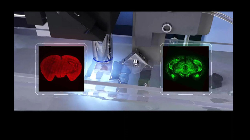
Sectioning-Imaging Mode
Innovative way to acquire whole brain imaging at single neuron level
Combine ultra-thin sectioning and high-contrast imaging in a unique,high-throughput way.
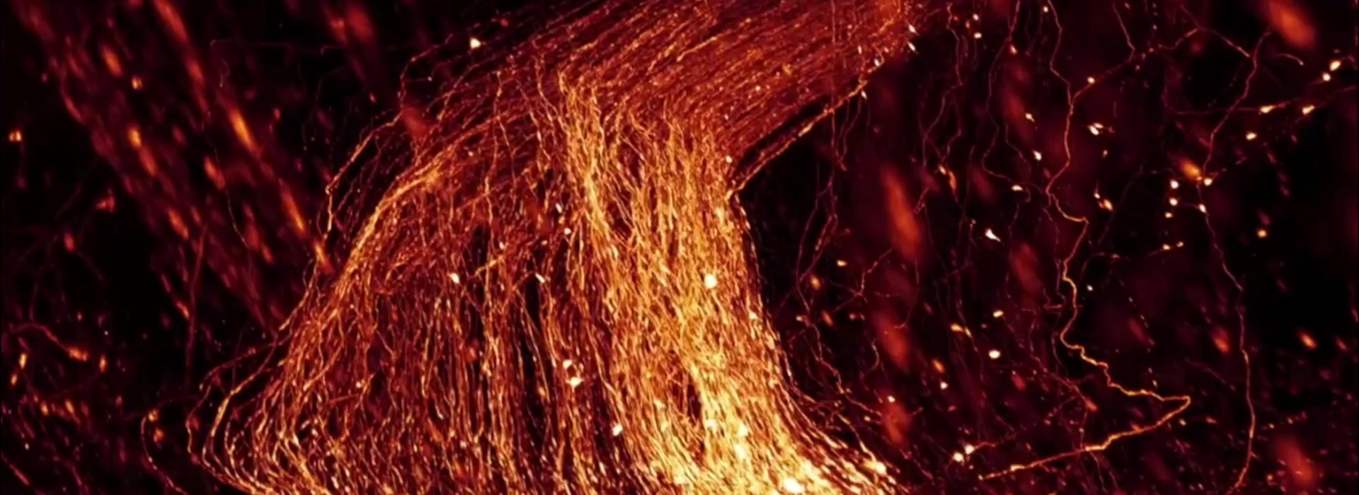
You are also able to enable visualization of synaptic-connection-related dendritic spines and axon boutons over the brain wide scale at the complete single neuron level.

Isotropic resolution
Demonstrate 3D data in any direction
Unlock isotropic resolution throughout the entire specimen.Self-registration of the whole-brain imaging data facilitates reconstruction of the whole tissue morphology in the most authentic way in 3D.

Resin embedding
Sample preparation made easy
Our optimised resin embedding method maintains the original shape of sample, preserves and enhances the fluorescence signal. The method can easily applied to various sample types including intact mouse, ferret and macaque brains as well as other organs.
You can also combine this method with other raditional staining methods such as Golgi, Nissl and HE staining.

Real-time staining
Rapid staining to maximize performance and efficiency
Simultaneously acquire labelled neural structures and cytoarchitecture reference using a real-time staining approach. This facilitates precise tracing of long-range projections and accurate locating of individual neurons in the whole brain.


Protein & particle distribution

