Technology
Technology
BioMapping series systems are based on Micro-Optical Sectioning Tomography (MOST) technology.
MOST technology enabless easy sample preparation, high image resolution & fast data acquisition.
Our unique and patented combination of sample sectioning and optical imaging enables neuroscientists worldwide to easily investigate whole-brain neuron projections and establish a standardized anatomical atlas.


Micro-Optical Sectioning Tomography (MOST)
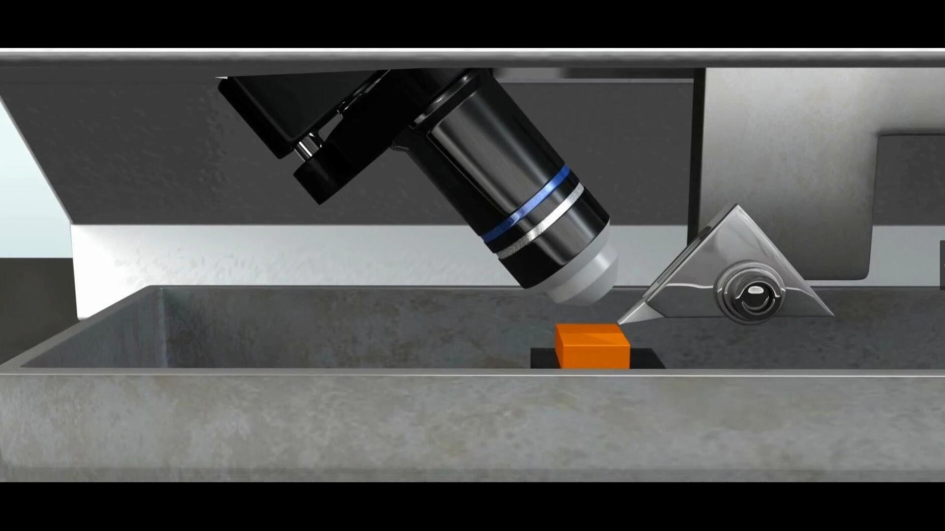
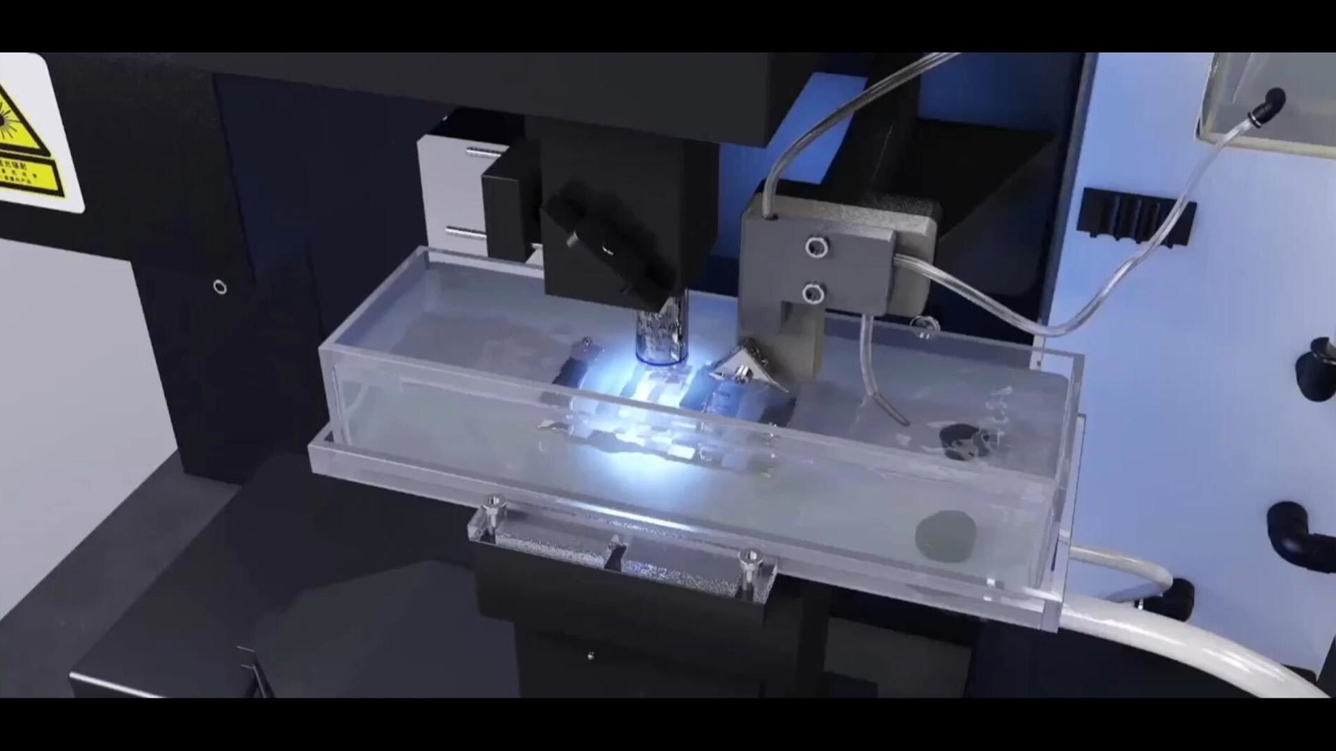
The MOST system is composed of a microtome, light microscope, and image recorder.
Our system performs imaging and sectioning simultaneously. During this process the microtome slices the specimen into sections.
After these sections have been removed from the rest of the sample, imaging takes place immediately. The light microscope is a reflected bright-field microscope in which the illumination beam is perpendicular to the rake face of the knife and coincides with the imaging beam.
As the sections are being imaged in motion, a time-delay integration line-scan charge-coupled device (TDI-CCD) is used to improve the sensitivity of image recording.
These design features, especially the optical design, can reduce system complexity to ensure automation stability and high resolution during time-consuming data acquisition without interruption.


fluorescence Micro-Optical Sectioning Tomography (fMOST)
Multi-channel laser lights initially pass through a series of optical components to generate a line spot. This line spot is focused on the sample surface to perform strip-by-strip scanning.
After excitation, the emission signal is recieved by the objective lens and captured by TDI-CCD cameras simultaneously.
All strip images captured can be precisely stitched to form an intact coronal section image. The surface of the sample is cut into ultra-thin slices to expose the new surface for imaging. The imaging-sectioning cycle repeats until the whole sample has been image.


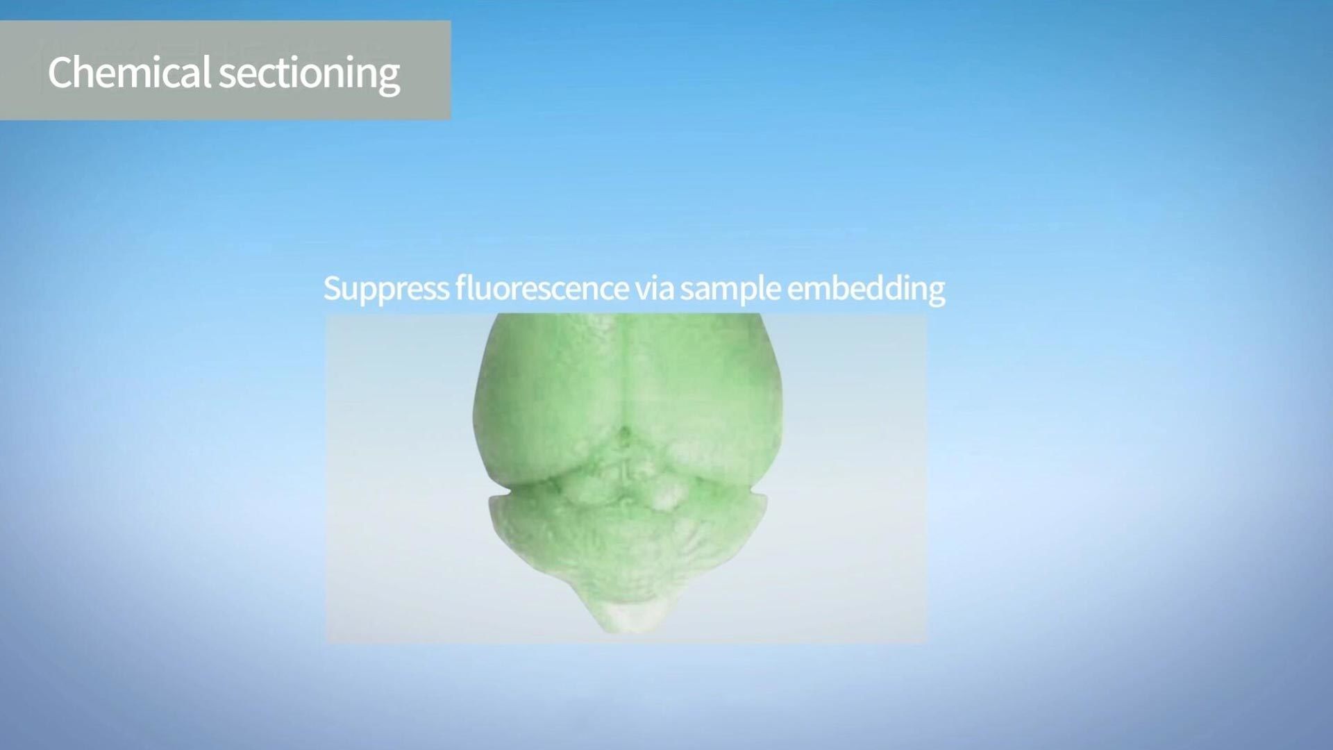
Chemical Sectioning
Our technology offers high-resolution axial sectioning. This is due to a fluorescence signal in the sample which can be switched to an ON & OFF state to avoid the out-of-focus background from deep layers.
During acidified resin embedding, the fluorescence in the brain tissue can be switched to a nonfluorescent state. Then, the fluorescence on the superficial layer of the trimmed block is chemically recovered to the fluorescent state by subsequent alkaline buffer reactivation.
You could then use fMOST imaging to image the recovered signal.
Once imaging is complete, the block surface is removed. This exposes the new layer which can be fluorescently recovered for imaging again. The cycles repeat until complete imaging of the intact tissue has been completed.


Real-time Staining
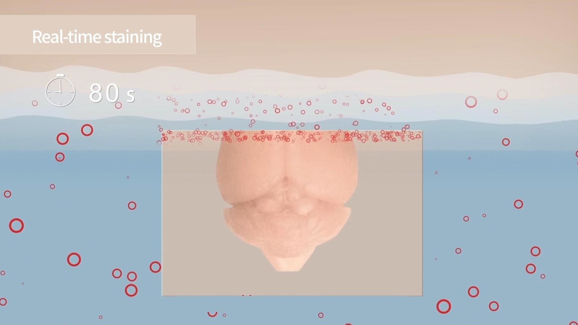
Many fast-staining dyes can be easily integrated into fMOST imaging. For example, propidium iodide (PI) solution can serve as the sample immersion liquid for imaging to achieve whole-brain counterstaining.
After sectioning, the dye immediately penetrates the fresh surface, combining with nucleic acid inside cell bodies and staining the soma, proximal dendrites and axon hillocks . Real time staining can be integrated with the fMOST imaging process. This means that experiments can be automatically conducted in continuous in-situ staining/imaging/sectioning cycles.
The technology uses a recirculating filtration device that can be applied to maintain a flattened section.This removes the cutting chips, purifies the PI solution. The recirculating filtration device helps to maintain a uniform PI concentration.
